





The 2026 Nobel Prize in Physiology or Medicine went to Karl Deisseroth, Peter Hegemann and Georg Nagel for optogenetics. Here is how an algal light sensor became a brain switch, what it has shown so far, and who was left off the podium.
Karl Deisseroth missed the call. It came in at 12:27 a.m. on Monday, October 5, and a second call, this one to his wife, the neuroscientist Michelle Monje-Deisseroth, got through from Stockholm. By morning he was one of three people holding the 2026 Nobel Prize in Physiology or Medicine.
The prize is shared with Peter Hegemann of Humboldt University in Berlin and Georg Nagel of the University of Würzburg, and it rewards research into how the brain switches individual nerve cells on and off. The short name for the toolkit is optogenetics. The long version involves a swimming alga, a seven-pass membrane protein, a viral vector, and a fiber thinner than a hair.
What follows covers the protein, the hardware, what the method has revealed, the one human trial that has taken it into a clinic, and the argument over who the committee left off the podium.
The Call Nobody Picked Up, and the Reel That Followed
Stanford posted a short video that same Monday, and it became the human face of the announcement. Deisseroth stands in jeans and a T-shirt, his wife behind him, and tells his daughters he got a call that morning about the prize. His son, still in pajamas, hears it next. One daughter handles the plot twist before anyone else can: as Boston.com transcribed it, “He’s sharing it with two other people.”
If the embed below does not load on your device, the reel lives on Stanford’s Instagram account.
Background worth having: Deisseroth was born in Boston in 1971, graduated from Harvard in 1992, then earned a PhD in neuroscience and an MD at Stanford, where he started his lab in 2004. He still practices psychiatry, treating treatment-resistant depression and autism spectrum disorders. Stanford counts him as its 37th Nobel laureate, and the full account is on Stanford Report.
What the Prize Actually Rewards: Control, Not Just Observation
Neuroscience spent most of the twentieth century listening. Electrodes recorded spikes, scanners mapped blood flow, and researchers inferred which cells mattered from what lit up. Correlation was the currency. Causation was harder to buy.
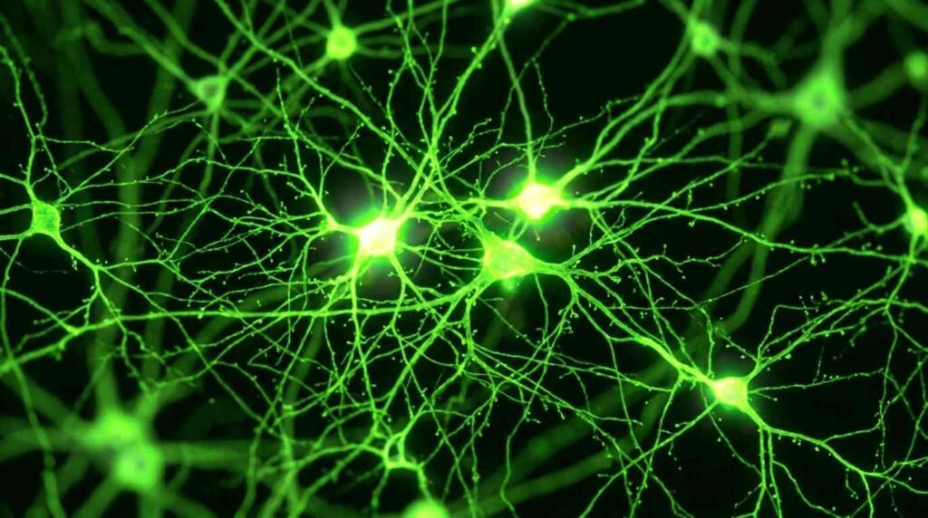
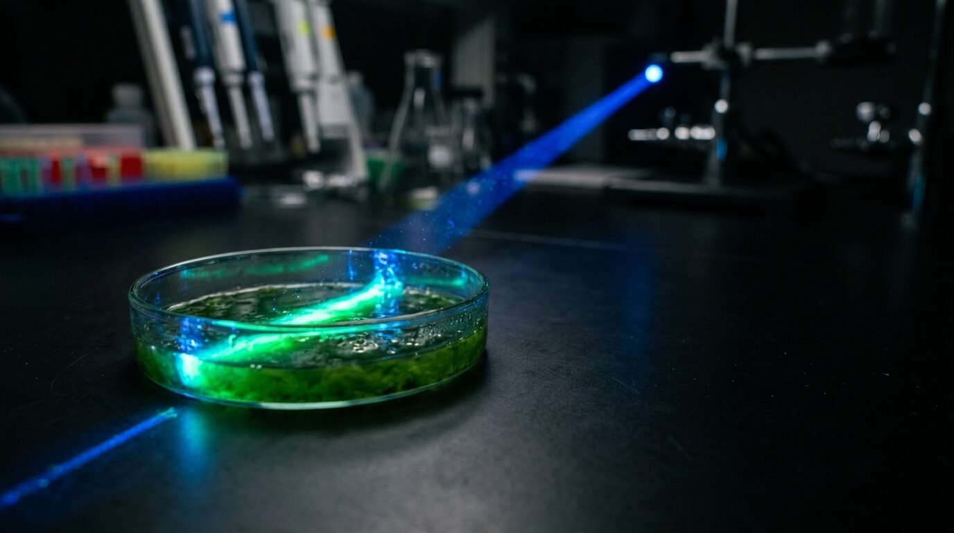
Optogenetics flips the direction of the experiment. Instead of watching a circuit, you reach in and drive it. Deisseroth framed the contrast to STAT in a single clause: “we’re using light to cause things to happen.” In practice, a researcher picks a cell type, makes only those cells light-responsive, and flashes light on a behaving animal while watching what changes.
The Nobel committee’s chair, Per Svenningsson, said the approach offers a way of mapping the brain “in a way that we could once only dream of,” as Nature reported. Rui Costa of the Allen Institute told the same outlet that it fundamentally changed how neuroscience is done.
The Protein: How a Swimming Alga Became a Switch
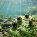
The story starts with a question that had nothing to do with brains. Hegemann, working in the early 1990s at the Max Planck Institute, wanted to know how the single-celled alga Chlamydomonas reinhardtii steers toward light. With Nagel he found the answer, a light-sensitive ion channel they named channelrhodopsin, and per STAT it opened rapidly when light hit it.
The design is what made it useful. According to Neuroscience News, channelrhodopsin is a single seven-transmembrane protein that works as both the light sensor and the ion pore, with no intermediate signaling cascade. Blue light opens the pore within milliseconds, and positively charged ions, mainly sodium and calcium, rush in. In a neuron, that inward current is an action potential waiting to happen.
Nagel and colleagues published the characterization in Science in 2002 and in PNAS in 2003. The decisive property turned out to be portability: the team found the protein worked in any cell into which it was introduced. Add the gene to a neuron, and the neuron answers to light.
Not from the sources above, so verify before relying on it: channelrhodopsins use a retinal cofactor, the same light-absorbing molecule found in the human eye’s rhodopsin, and the commonly used version responds best to blue light near 470 nanometers. Silencing tools, which pump ions out or let negative ions in, were developed afterward and complete the on/off pair.
The Hardware: Vectors, Promoters and a Fiber the Width of a Hair
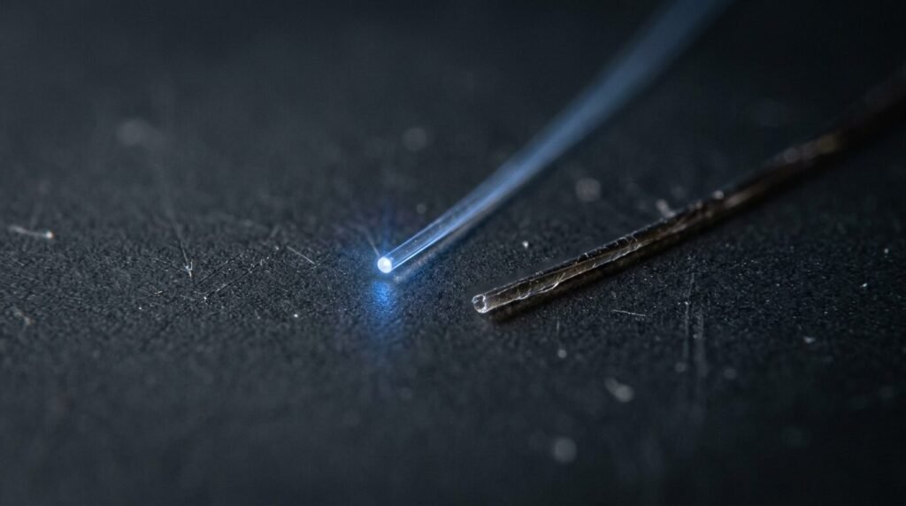
A protein from an alga is a curiosity until you can deliver it and trigger it. Two milestones turned it into an instrument. In 2005, Deisseroth’s team delivered the channelrhodopsin-2 gene into cultured rat neurons using viral vectors, and brief flashes of blue light produced synchronized action potentials, per Neuroscience News. In 2007, the tool moved into living, awake, freely moving animals, with cell-type-specific promoters and hair-thin fiber-optic implants.
Each layer of that rig solves a different problem, and each carries its own constraint:
| Component | Job | Practical limit |
|---|---|---|
| Opsin gene (e.g., channelrhodopsin-2) | Makes a cell light-responsive | Must be expressed in the right cells at the right level |
| Viral vector (typically AAV) | Carries the gene into tissue | Delivery requires an injection; packaging capacity is finite |
| Cell-type-specific promoter | Restricts expression to chosen neurons | Specificity depends on the promoter and the target region |
| Fiber-optic implant | Delivers light deep in tissue | Invasive in animals; tissue scatters light |
| Light source (laser or LED) | Triggers the channel in milliseconds | Intensity and heating must be controlled |
The table’s “practical limit” column is a general summary rather than a sourced list. The Neuroscience News piece implies the constraints, since it notes that delivery requires genetic vectors and that in vivo use required implanted fiber optics, but it does not discuss limitations directly.
What It Has Shown About the Brain So Far
Once you can command a cell type on demand, whole categories of old questions become experiments. The same Neuroscience News summary lists several. Stimulating or silencing the projections among the ventral tegmental area, the nucleus accumbens, and the prefrontal cortex has been used to probe circuits linked to anhedonia in depression, compulsive drug seeking, and panic in PTSD.
In Parkinson’s disease models, the tool clarified how the brain’s direct and indirect basal ganglia pathways interact, which the article says helps explain why deep brain stimulation works. And in memory research, groups tagged the sparse ensembles of neurons encoding a particular memory, then showed that light could reactivate, modify, or erase traumatic recall in animals.
For Deisseroth the clinical relevance runs through explanation. Speaking with WBUR, he argued that psychiatry needs a biological account of why patients suffer, and that once the relevant cells are identified, “you can design any kind of treatment.”
From Mice to a Patient: The Vision Trial
The most concrete human result so far came from the eye. In 2021, Nature Medicine published a case from the groups of Botond Roska in Basel and José-Alain Sahel at the University of Pittsburgh, a collaboration GEN describes as 13 years long. The patient was a 58-year-old man with retinitis pigmentosa, blind after his photoreceptors degenerated.
The team injected an AAV vector carrying ChrimsonR, an optogenetic sensor, into one eye, targeting the retinal ganglion cells that still survived. Because those cells are not naturally light-sensitive, he wore engineered goggles with a camera that converted the scene into light pulses projected onto the retina in real time, at a wavelength matched to the opsin.
Before treatment, the goggles gave him nothing. Afterward he could recognize, count, locate, and touch objects on a table. Sahel said he spontaneously reported seeing “some stripes, black and white stripes on the street.” Visual behavior also tracked with brain activation associated with vision.
The caveats are large, and the sources say so. It was one individual in an ongoing Phase I/IIa trial, the recovered vision was partial, and Bainbridge of University College London cautioned that more evidence would be needed to judge whether the technology provides useful vision. Treat it as a proof of principle, not a cure.
Who Was Left Off the Podium
Nobel rules cap a prize at three people, and optogenetics has more than three claimants. STAT reported that Ed Boyden, first author on the landmark 2005 paper while a PhD student in Deisseroth’s lab and now at MIT, was not named, and that many commentators on social media said he should have been. Boyden did not respond to STAT’s request for comment, and the committee chair declined to explain the selection.

Others appear in the Nobel background material, per the same article, including Gero Miesenböck in 2002 and Zhuo-Hua Pan in 2004, whose work in retinal ganglion cells was submitted late that year but not published until 2006, after the Deisseroth and Boyden paper had landed. How credit should be sliced in a field built by many hands is an old argument, and this prize reopens it without settling anything.
The same week produced two other science prizes covered on this site: the physics award for the IceCube neutrino detector and the chemistry award for one-handed molecules.
Questions Readers Are Asking
Is optogenetics a treatment today? Not in general. It is overwhelmingly a research method. The retinitis pigmentosa case is the clearest clinical use, and it remains an early trial.
Does it require implants in people? The animal work relied on implanted fibers. The vision case instead used external goggles, because the retina is already exposed to light.
How did Deisseroth himself explain it? In a Stanford video relayed by ABC News, he put it this way: “Optogenetics is a way of turning cells on or off with light.”
How much is the prize worth? Twelve million Swedish kronor, about $1.2 million, divided equally among the three laureates.
Sources: NobelPrize.org, Nature, STAT, Neuroscience News, WBUR, Boston.com, ABC News via 6abc, GEN, and Stanford Report.


One thought on “A Flick of Blue Light, a Nobel Prize: How Optogenetics Turned an Algal Protein Into a Brain Switch”